
A small rough area on the edge of the tongue, a white spot that doesn’t go away after two weeks, a diffuse pain while eating. These situations lead many people to search for images online to compare. However, tongue cancer remains difficult to identify visually without a medical examination, as several benign lesions resemble it closely.
HPV Status and Tongue Cancer: A Data Point That Changes Prognosis
Content about tongue cancer often mentions tobacco and alcohol as the main risk factors. The role of human papillomavirus (HPV) deserves particular attention, especially for tumors located at the base of the tongue.
HPV 16 is the strain most frequently associated with oropharyngeal cancers. These tumors affect a different patient profile: often younger, without excessive tobacco or alcohol consumption. According to an anatomical-pathological review published on MSDConnect incorporating WHO recommendations 2022-2023, HPV-associated tumors have a significantly better prognosis than HPV-negative tumors, with significantly higher overall survival rates.
This difference has concrete consequences. Since the revision of the TNM classifications for the oropharynx, HPV status (detected by immunohistochemistry p16) officially modifies the tumor stage. It also guides therapeutic de-escalation strategies, meaning less intensive treatments, with fewer effects on speech and swallowing. Vaccination against HPV, recommended in France for both girls and boys, serves as a direct preventive measure against these cancers.
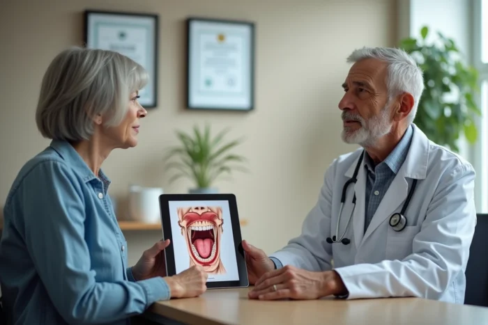
When trying to identify a problem on your tongue, it may be helpful to consult photos of cancer or white spots on the tongue to better understand the visual differences between lesions, although no image can replace medical advice.

White Spot on the Tongue: Three Diagnoses to Distinguish
Have you noticed a whitish patch on your tongue? Before thinking about cancer, know that the vast majority of these spots correspond to benign conditions. Three diagnoses account for almost all cases.
Leukoplakia
Leukoplakia appears as a white patch, sometimes slightly raised, that does not detach when scraped. It is the only one of the three that can progress to cancer. Two forms are distinguished:
- Homogeneous leukoplakia: regular, white, and flat surface. Its risk of malignant transformation remains low.
- Non-homogeneous leukoplakia: irregular surface, mixing white and red areas (this is referred to as erythroleukoplakia). The risk of cancerous transformation is higher for this form.
- In both cases, only a biopsy can confirm the exact nature of the lesion. Visual appearance alone is not sufficient to decide.
Oral Thrush and Coated Tongue
Thrush (oral candidiasis) forms a creamy white deposit that can be removed by rubbing, leaving a red mucosa underneath. It often affects people on antibiotics, immunocompromised individuals, or those wearing braces.
The coated tongue, on the other hand, is simply a whitish coating related to poor oral hygiene, dehydration, or gastric reflux. It disappears with gentle brushing of the tongue. Neither thrush nor coated tongue are precancerous lesions.
Clinical Signs of Tongue Cancer Beyond Color
A white spot is just one sign among others. Squamous cell carcinoma, which represents the vast majority of tongue cancers, also manifests through symptoms that a photo cannot show.
- A persistent ulceration on the lateral edge or underside of the tongue that does not heal after two to three weeks despite local care.
- A unilateral pain radiating to the ear on the same side, with no identified ENT cause.
- An indurated area (hard to the touch), even without pain. This thickening beneath the mucosa is a signal that photos cannot detect.
- A palpable cervical lymph node, firm and non-painful, present for several weeks.
- Difficulties in sticking out the tongue or moving it normally, a sign of muscular infiltration.
Persistence beyond three weeks is the main triage criterion. An aphthous ulcer heals spontaneously. A cancerous lesion does not.
Urgent Consultation or Scheduled Appointment: Triage Criteria
Not all tongue lesions require the same degree of urgency. Knowing how to differentiate helps avoid both diagnostic delays and unnecessary visits to the emergency room.
Consultation with a Doctor or Dentist Within Two Weeks
An isolated white spot, without bleeding or intense pain, justifies a quick consultation but not a hospital emergency. The practitioner will evaluate the lesion, may request a biopsy, and refer to an ENT specialist or a stomatologist if necessary.
Emergency Room Visit or Call to 15
Some situations require immediate attention: heavy bleeding from the tongue that does not stop, sudden difficulty swallowing or breathing, or rapid swelling of the floor of the mouth. These symptoms may indicate a large tumor compressing the airways.
Risk Factors for Tongue Cancer: Beyond Tobacco
Tobacco remains the best-documented risk factor. It multiplies the risk of carcinoma of the oral cavity. The tobacco-alcohol combination further amplifies this risk, as both substances act synergistically on the mucosal cells.
Other factors deserve attention. Chronic trauma (a broken tooth rubbing against the tongue, poorly fitting dentures) creates repeated local irritation. Prolonged poor oral hygiene also increases the risk. HPV, mentioned earlier, mainly concerns the base of the tongue and the oropharynx.
The diagnosis always relies on a biopsy, supplemented by imaging studies (CT scan, MRI) to assess the extent of the tumor and determine the stage. Neither an online photo nor a self-examination in front of the mirror can replace this examination. In the face of a persistent lesion on the tongue, the only reliable course of action is to consult a healthcare professional who can palpate the area, check the cervical lymph nodes, and decide on further examinations.